filmov
tv
microscopy image
0:02:01
Extracellular Vesicles (EVs) Imaging by Atomic Force Microscopy | Protocol Preview
0:04:08
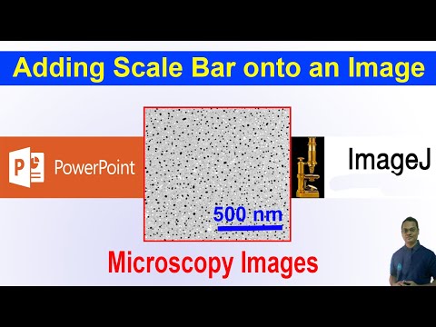
Adding a Scale Bar onto a Microscopy Image using PowerPoint/ImageJ | Drawing/Graphing-09
0:00:24
Latest Image of An Atom! 🔬
0:00:06
Light sheet microscopy image of zebrafish in vivo imaging
0:29:29
Meghan Driscoll - Analyzing morphological motifs in 3D microscopy images
0:00:19
Green apple A Hidden World Under the Microscope – Bacteria Close-Up #microscope #science #biology
0:00:33
Light sheet microscopy image of pancreatic carcinoma cell line with infiltrating CAR T cells
0:00:35
Can You Guess What This Microscope Image Is?
0:01:03
Cell Counting Made Easy: Using ImageJ to Analyze Microscopy Images
0:05:42
Two-photon Microscopy and Calcium Imaging. Tools to study the functioning brain
1:14:50
Artificial Intelligence and Histopathological Characterization of Microscopy Images
0:00:28
Eye-Trace: Segmentation of Volumetric Microscopy Images with Eyegaze
0:33:41
Microscopy: Cameras and Digital Image Analysis (Nico Stuurman)
0:00:21
Lipstick under the microscope A Hidden World Under the Microscope – Bacteria Close-Up!
0:01:00
What's The Smallest You Can See?
0:03:13
Image-Based Autofocus in Microscopy
1:25:54
Complete and Fast 3D Image Analysis in Microscopy
0:00:29
Light sheet microscopy image of identifying tumor target sites for effective therapies
0:00:15
AI generated Scanning Electron Microscope image zoom.
0:22:44
Microscopy and Imaging Center Virtual Tour
0:28:51
A FAIR Share: Annotate Microscopy Image Data with Curated Vocabularies
0:17:42
David Albrecht: Nanoscale imaging of live cells with confocal interferometric scattering microscopy
1:09:27
Markus Sauer - Super-resolution expansion microscopy - Imaging ONEWORLD
0:31:10
Discovering transport of RNA skeletal muscle 3D microscopy image analysis & Markov| SciPy 2021
Назад
Вперёд
 0:02:01
 0:04:08
 0:00:24
 0:00:06
 0:29:29
 0:00:19
 0:00:33
 0:00:35
 0:01:03
 0:05:42
 1:14:50
 0:00:28
 0:33:41
 0:00:21
 0:01:00
 0:03:13
 1:25:54
 0:00:29
 0:00:15
 0:22:44
 0:28:51
 0:17:42
 1:09:27
 0:31:10